Hip And Upper Thigh Anatomy / Right Upper Thigh Hip Pain - You can ask your partner to abduct his or her thigh to feel for contraction of gluteus medius and gluteus minimus.
byAdmin-
0
Hip And Upper Thigh Anatomy / Right Upper Thigh Hip Pain - You can ask your partner to abduct his or her thigh to feel for contraction of gluteus medius and gluteus minimus.. Twists the leg out and twists the knee in toward your other leg. The uppermost of the medial thigh muscles is the pectineus muscle. The anatomical areas found on the upper limb can serve as key landmarks to help us find important anatomical structures such as finding one of the superficial veins: Anatomy hip, thigh and leg muscles. Muscle origins (o) are shown in red, insertions (i) in blue.
Groin, inguinal region and the anterior and posterior regions of the hip and thigh. B, muscles of the anterior thigh compartment. A, anterior and posterior views show the hip joint ligaments. The median cubital vein (a common site site for venepuncture) in the antecubital fossa of the arm. There are a lot of muscles of the hip and thigh.
The Hip Joint Joints Seam from sites.google.com Popular study materials from anatomy and cell biology 306. The different anatomical areas of the gluteal region: The information contained in anatomy atlases is not a substitute for the medical care and advice of your physician. Knee assessment and hip mechanics online course: 3d interactive models and video tutorials on the anatomy of the thigh, including musculature, bones, blood supply and innervation. He also serves the communities of charleston, sc and augusta, ga. The ilium, the i was toks5b saw rxray upper hip us dislocated from pelvic n laying assuced n directly on top of the lower hip. Pelvis, perineum, hip, and upper thigh.
The upper part of the thigh bone consists of the femoral head, femoral.
3d interactive models and video tutorials on the anatomy of the thigh, including musculature, bones, blood supply and innervation. Learn their anatomy efficiently and easily using kenhub's. Anatomy hip, thigh and leg muscles. Anterior view (i) right limb. Thigh muscles also protect neurovascular structures as they go through the proximal hip joint to the knee and lower leg(3). Want to learn more about it? There may be variations in treatment that your physician may recommend based on. This vein, as well as the deep veins. Groin, inguinal region and the anterior and posterior regions of the hip and thigh. Quadriceps, a group of four. Twists the leg out and twists the knee in toward your other leg. The upper part of the thigh bone consists of the femoral head, femoral. Bends (flexion) the thigh at the hip.
Study 14 hip/upper thigh muscles flashcards from colleen k. Learn their anatomy efficiently and easily using kenhub's. The information contained in anatomy atlases is not a substitute for the medical care and advice of your physician. The upper part of the thigh bone consists of the femoral head, femoral. Hip surgeon dr guillaume dumont offers hip pain treatments in columbia, sc.
The Hip Joint Joints Seam from sites.google.com It is inserted between the two layers of the iliotibial band of the fascia lata about the junction of the middle and upper thirds of the thigh. Pelvis, perineum, hip, and upper thigh. Groin, inguinal region and the anterior and posterior regions of the hip and thigh. Click on the link to view the high quality hip anatomy video provided by your practice online. The median cubital vein (a common site site for venepuncture) in the antecubital fossa of the arm. A, anterior and posterior views show the hip joint ligaments. The information contained in anatomy atlases is not a substitute for the medical care and advice of your physician. 24.14 muscles of the hip and thigh:
In vertebrate anatomy, hip (or coxa in medical terminology) refers to either an anatomical region or a joint.
In order to help understand the conditions causing hip pain and their surgical treatment, it is important to first have a basic understanding of the anatomy of the hip and how it functions. Anatomy hip, thigh and leg muscles. He also serves the communities of charleston, sc and augusta, ga. Hip anatomy human body anatomy human anatomy and physiology anatomy study leg muscles anatomy thigh muscle anatomy inner thigh hip pain explained. Study 14 hip/upper thigh muscles flashcards from colleen k. Atlas of human anatomy in cross section. Related online courses on physioplus. Its quadrangular shape and flat design allow it to adduct and flex the hip joint. Hip surgeon dr guillaume dumont offers hip pain treatments in columbia, sc. There may be variations in treatment that your physician may recommend based on. In vertebrate anatomy, hip (or coxa in medical terminology) refers to either an anatomical region or a joint. Work the small muscles of your inner thighs—often overlooked in yoga—to find ease in all sorts of poses. Ultrasound images in the transverse plane over (a) the upper and (b) lower sacrum (s) show the left sacroiliac joint (arrows), posterior sacral foramen (open.
Learn their anatomy efficiently and easily using kenhub's. There are a lot of muscles of the hip and thigh. Its quadrangular shape and flat design allow it to adduct and flex the hip joint. While the thigh muscles will be slip into the anterior, medial and posterior groups. 340 anatomical structures of the hip region were labeled, accessible on anatomical parts:
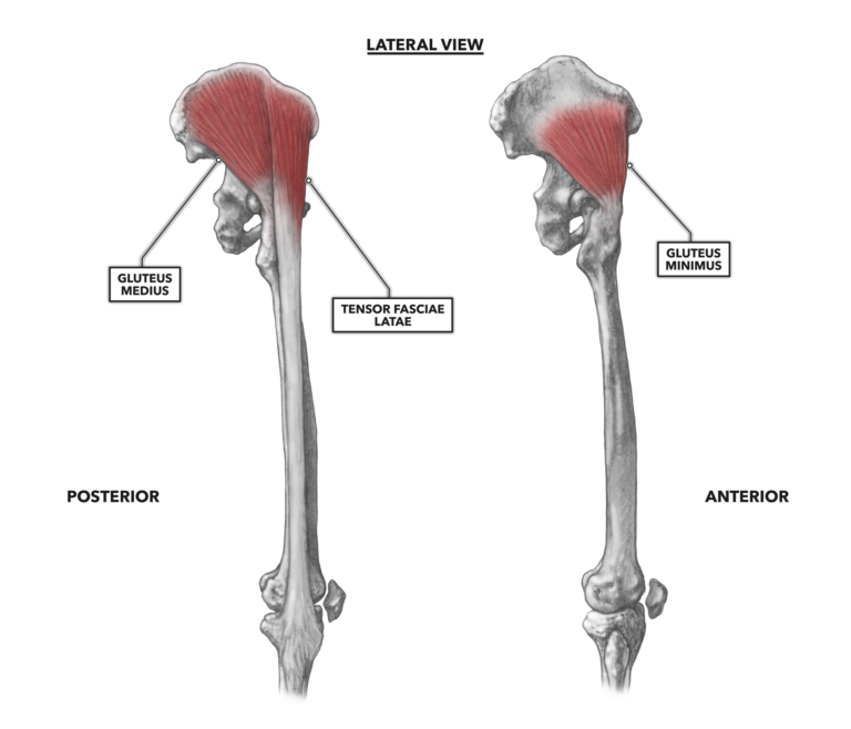
Crossfit Hip Musculature Part 3 Lateral Muscles from www.crossfit.com The upper part of the gluteus maximus muscle, and the gluteus medius muscle beneath. The anatomical areas found on the upper limb can serve as key landmarks to help us find important anatomical structures such as finding one of the superficial veins: Groin, inguinal region and the anterior and posterior regions of the hip and thigh. In some people with a significant lumbar curve, psoas major pulls on the upper lumbar vertebrae and t12 posteriorly. The cavity of the acetabulum faces obliquely forward, outward, and downward. The iliopsoas muscle, which extends from the lower back to upper femur; There may be variations in treatment that your physician may recommend based on. The femur, the hip bone (subdivided into ilium.
Click on the link to view the high quality hip anatomy video provided by your practice online.
The adductor muscle on the inner thigh; There may be variations in treatment that your physician may recommend based on. Local nerves running through and around the hip & pelvis. Sartorius muscle anatomy page has origin, insertion, innervation, and blood supply information. Hip surgeon dr guillaume dumont offers hip pain treatments in columbia, sc. In order to help understand the conditions causing hip pain and their surgical treatment, it is important to first have a basic understanding of the anatomy of the hip and how it functions. The ilium, the i was toks5b saw rxray upper hip us dislocated from pelvic n laying assuced n directly on top of the lower hip. Work the small muscles of your inner thighs—often overlooked in yoga—to find ease in all sorts of poses. Bends (flexion) the thigh at the hip. While the thigh muscles will be slip into the anterior, medial and posterior groups. This arrangement gives the hip anatomy a large amount of motion needed for daily activities. In vertebrate anatomy, hip (or coxa in medical terminology) refers to either an anatomical region or a joint. Groin, inguinal region and the anterior and posterior regions of the hip and thigh.